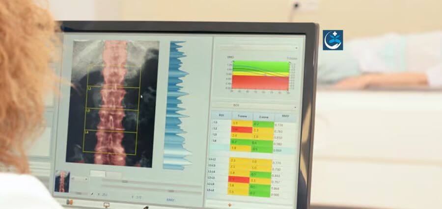
Machine learning has been used by researchers to evaluate bone density scans for calcification in the body’s main artery, the aorta. They claim that even before symptoms develop, their technology could be used to forecast future cardiovascular and other diseases.
The calcification of the aorta, the body’s biggest artery, can cause problems in the same way as calcium deposits on the inner walls of blood arteries in the heart can. After leaving the heart, it extends downward to the abdomen, where it divides into smaller arteries that supply each leg, and upward to serve the brain and arms.
Calcification in the portion of the aorta that passes through the abdomen known as abdominal aortic calcification (AAC) might predict the onset of cardiovascular disorders including heart attack and stroke as well as mortality risk. It is a valid marker for dementia in old age, according to earlier studies. AAC can be seen on bone density scans, which are frequently used to identify osteoporosis in the lumbar vertebrae, but it requires time and a highly qualified expert to analyse the results.
The 24-point AAC-24 rating system is frequently used by qualified imaging specialists to quantify AAC. The least severe form of AAC is represented by a score of zero, while the most severe is represented by a score of 24. In order to accelerate the calcification assessment and scoring process, researchers from Edith Cowan University in Australia are now using machine learning.
The researchers fed their machine learning model 5,012 spinal pictures from four distinct bone density machine models. The researchers claim that this study is the largest and the first to use images from standard bone density tests to test the algorithms in a real-world environment.

