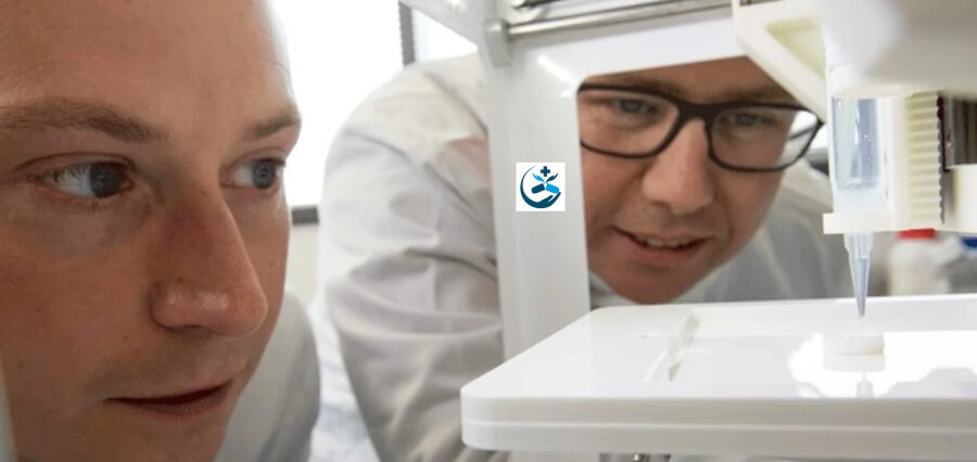
Using 3D photolithographic printing, a group of bioengineers and biomedical scientists from the Children’s Medical Research Institute (CMRI) at Westmead and the University of Sydney have developed a complicated environment for constructing tissue that resembles the architecture of an organ.
The teams were led by developmental biologist Professor Patrick Tam, head of the CMRI’s Embryology Research Unit, and professors Hala Zreiqat and Peter Newman from the University of Sydney’s School of Biomedical Engineering.
The technique was used to teach stem cells obtained from skin or blood cells to become specialised cells that can group together to form an organ-like structure using bioengineering and cell culture techniques.
Cells use carefully positioned proteins and mechanical triggers to travel through their complex environment and replicate developmental processes, much how the needle of a record player manipulates the grooves of the vinyl record to produce music. In their most recent study, the scientists mimicked cellular processes during growth using microscopic mechanical and chemical signals.
Professor Hala Zreiqat said: “Our new method serves as an instruction manual for cells, allowing them to create tissues that are better organised and more closely resemble their natural counterparts. This is an important step towards being able to 3-D print working tissue and organs.”
Similar to putting together a structure from many different pieces, Dr Newman stated that creating tissues from cells required comprehensive instructions: “Imagine trying to build a Lego castle by throwing the blocks randomly on a table and hoping that they’ll fall into the right spot. Despite the fact that each brick is intended to link with others, without a clear plan, the result would probably resemble a big collection of broken Lego blocks rather than a castle.
| Read More: Click Here |


